


SiPuNai Medical | Validation Study of an AI Automatic Lenke Classification Model for Adolescent Idiopathic Scoliosis: A Literature-Based Analysis
SiPuNai Official Account

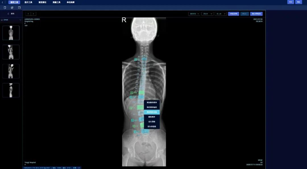
Recently, the team of Prof. Zhao Changqing, Department of Orthopedics, Ninth People's Hospital, Shanghai Jiao Tong University School of Medicinepublished a research paper in the Journal of Spinal Surgeryentitled "Validation Study of an AI Automatic Lenke Classification Model for Adolescent Idiopathic Scoliosis". This study used SiPuNai Medical Technology (Shanghai) Co., Ltd.developed the Ruiji (SpineSight) image processing software to build an AI model for measuring AIS patients' Cobb angles and performing Lenke classification, evaluating its feasibility and practicality in clinical practice.


This study developed an AI model to automatically measure the Cobb angle of adolescent idiopathic scoliosis (AIS) and perform Lenke classification, aiming to improve measurement efficiency and consistency. Although AI methods (e.g., CNN) achieve acceptable consistency with manual measurements, clinical adoption still faces challenges such as "black-box diagnosis" and medical liability. The study emphasizes that enhancing algorithm interpretability and allowing manual verification and adjustment can increase clinicians' trust in automated measurement and promote its application in practice.
1. Literature Search
Inclusion criteria: 1) clear AIS diagnosis with definite Lenke classification; 2) patient age 10–20 years; 3) clear and complete imaging data.
Exclusion criteria: 1) poor image acquisition or stitching quality; 2) severe pelvic tilt; 3) previous spinal surgery history.
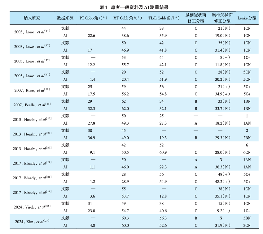
Based on the above criteria, 7 publications with 14 cases were finally selected (1 male, 13 female; age 14.88±2.55 years). All imaging data were used as the validation set to evaluate the accuracy of Cobb angle measurement and Lenke classification.
2. Model Construction
2.1. Landmark Detection
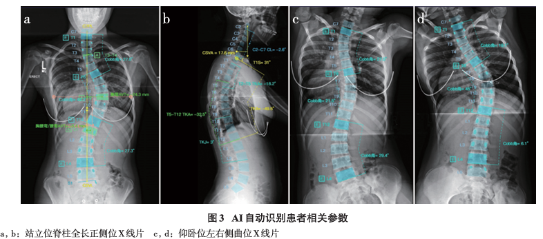
Based on the Ruiji (SpineSight) image processing software, this study developed an AI model for automatically measuring spinal parameters. The model uses an asymmetric encoder-decoder network architecture. The encoder is based on the RepVGG-A1 architecture, which introduces ResNet-like shortcut connections during training to ensure smooth gradient propagation, while being equivalent to an efficient VGG-style convolutional network at inference. With approximately 13M parameters, the model balances powerful feature extraction with real-time inference speed.
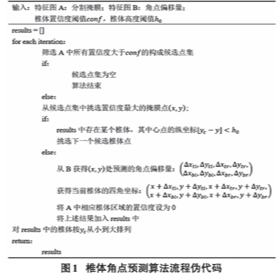
The model performs two key tasks: generating pixel-wise segmentation masks of the vertebrae and predicting the offset between each vertebral pixel and its four corner landmarks. During training, the segmentation task is supervised with binary cross-entropy loss, while the corner regression task is optimized with smooth L1 loss. At inference, the algorithm combines the segmentation mask and predicted offsets to compute precise vertebral corner coordinates.
Performance validation showed that the model's mean radial error (MRE) in locating vertebral landmarks on standing full-length spine X-rays was only 1.24 mm on anteroposterior films and 1.28 mm on lateral films, with successful detection rates (SDR) within 2 mm reaching as high as 90.46% and 88.93%, respectively.In addition, the Dice similarity coefficient (DSC) reached 0.83 for pedicle segmentation, further confirming the model's high accuracy and clinical practicality in spinal image analysis.
2.2. Cobb Angle Measurement
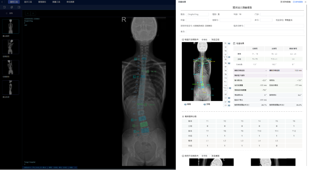
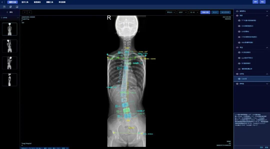
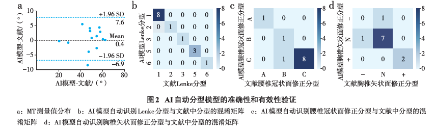
This study Lenke classificationdetermined the Cobb angle measurement regions based on Lenke classification, identified vertebral landmarks on full-length anteroposterior and lateral bending X-rays with the AI model, and automatically calculated the Cobb angle of each region. Bland-Altman analysis was used to evaluate agreement. Results showed excellent agreement between automated and manual measurements (ICC>0.86), with small mean absolute error and root mean square error for the major curve Cobb angle, and measurement differences within the clinically acceptable range of 5°, demonstrating the reliability and practicality of the automated method in classification and measurement.
2.3.Lenke classification
This study used the AI model to automatically measure Cobb angles and sagittal parameters, performing Lenke main classification and modifier classifications of lumbar coronal (A/B/C) and thoracic sagittal (-/N/+) types. Validation on external datasets showed the model achieved accuracies of 89%, 89%, and 96% on the three classifications respectively, confirming the high accuracy of the automated classification system and providing a reliable tool for clinical use.
3. Statistical Analysis
Statistical analysis was performed using SPSS, GraphPad Prism, and PyCharm software. After Shapiro-Wilk normality testing, measurement data were expressed as mean ± standard deviation, and between-group comparisons were made with t-tests. Reliability was assessed using the intraclass correlation coefficient, absolute agreement was measured by mean absolute difference and its standard deviation, and Bland-Altman plots were used to analyze agreement and systematic bias. The classification performance of the AI model was evaluated using confusion matrix metrics (accuracy, precision, recall, etc.). Differences were considered statistically significant at P<0.05.
The performance of the AI model was evaluated through external validation on data from 14 patients in the literature (covering Lenke types 1, 2, 3, 5, and 6). Results showed that the AI model's automatic measurements of the main thoracic curve (MT) (20.4°–62.09°) showed no significant difference from the literature values (20°–62°) (P>0.05), with excellent reliability (ICC=0.95). Bland-Altman analysis confirmed the good agreement between the two.
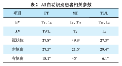
Regarding classification, the AI model achieved 100% accuracy on Lenke main classification. For lumbar coronal modifier (A/B/C) and thoracic sagittal modifier (-/N/+), the accuracies were 90.91% and 81.82%, respectively. Case-by-case validation further showed that the AI model's recognition of end vertebrae and apical vertebrae and its Cobb angle measurement for each curve type were fully consistent with the literature results.
Most notably, the model demonstrated extremely high efficiency, requiring only about 25 seconds from image import to complete measurement results and classification, far faster than the 30–400 seconds of manual measurement, fully demonstrating its great potential to improve diagnostic efficiency in clinical practice.
This study validated the application value of an AI-based automated model in the diagnosis of adolescent idiopathic scoliosis (AIS). By precisely locating vertebral corners with landmark detection technology, the model achieved automatic Cobb angle measurement and Lenke classification.
Results showed that the AI model's Cobb angle measurements of the main thoracic curve (MT) were highly consistent with literature data, with an intraclass correlation coefficient (ICC) of 0.95, demonstrating excellent measurement reliability. For classification, the model achieved 100% accuracy on Lenke main classification, 90.91% on lumbar coronal modifier, and 81.82% on thoracic sagittal modifier. Its efficiency advantage is particularly prominent: completing the full measurement and classification process in about 25 seconds, significantly faster than manual measurement.
Although the model maintained high accuracy when facing challenges such as image quality variations and anatomical overlap in the upper thoracic region, the study also noted limitations, including the limited sample size of the validation set and its failure to cover all rare scoliosis subtypes, which limits the model's generalizability.
In conclusion, this AI model provides a reliable tool for precise and efficient diagnosis of AIS and is expected to optimize clinical workflows and assist treatment decisions in future practice. Further research with larger samples and more comprehensive disease coverage is needed to enhance its generalizability.
SPINEX Spinal Holographic Intelligent Diagnosis & Treatment Platform